Quantitative in vivo corneal and ocular microscopy
The group “Quantitative in vivo corneal and ocular microscopy” led by Kristina Irsch focuses on solving problems inherent to ophthalmic imaging in those suffering from actual pathology. The overall goal is to realize the potential of the cornea and the eye to serve as a quantitative in vivo microscope into ocular as well as systemic diseases.

Presentation
The group develops innovative tools using original physics-based concepts, with two lines of research guiding its work :
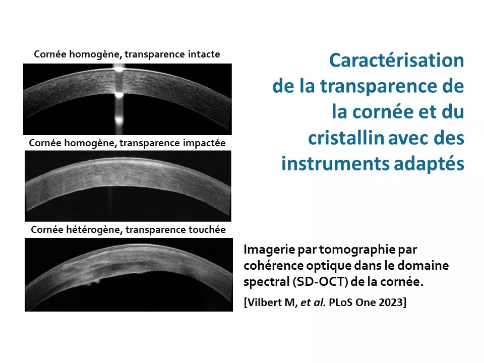
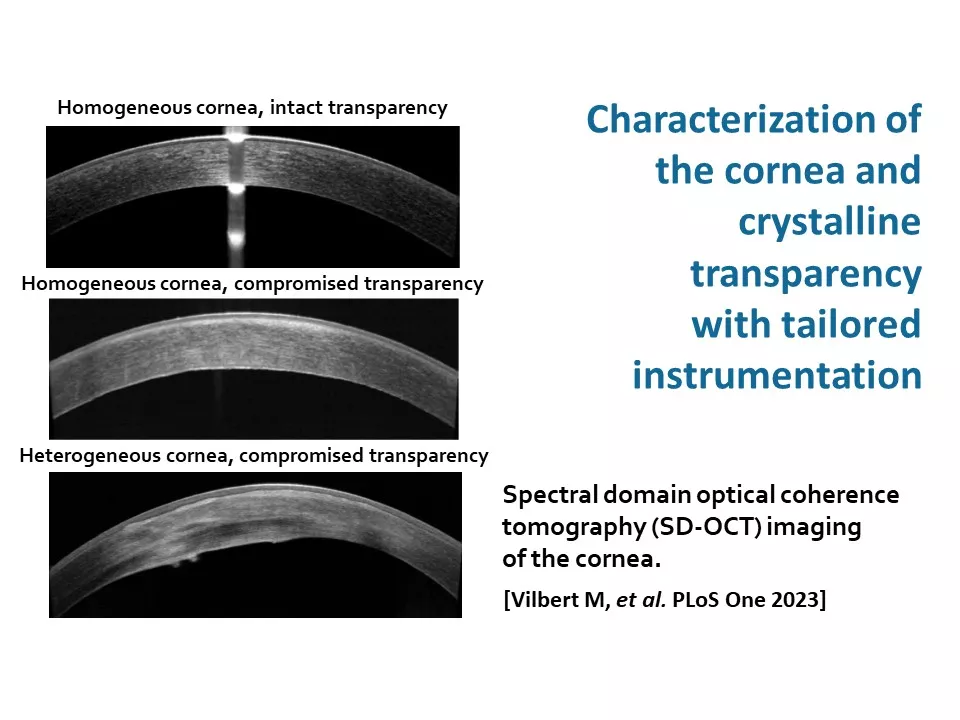
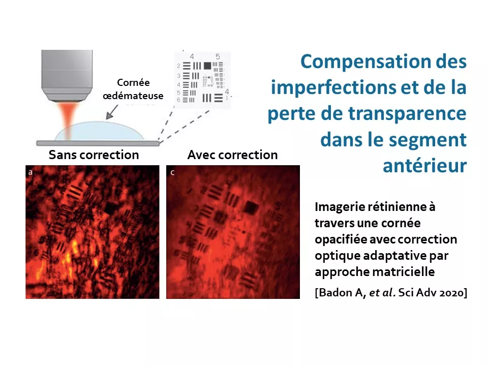
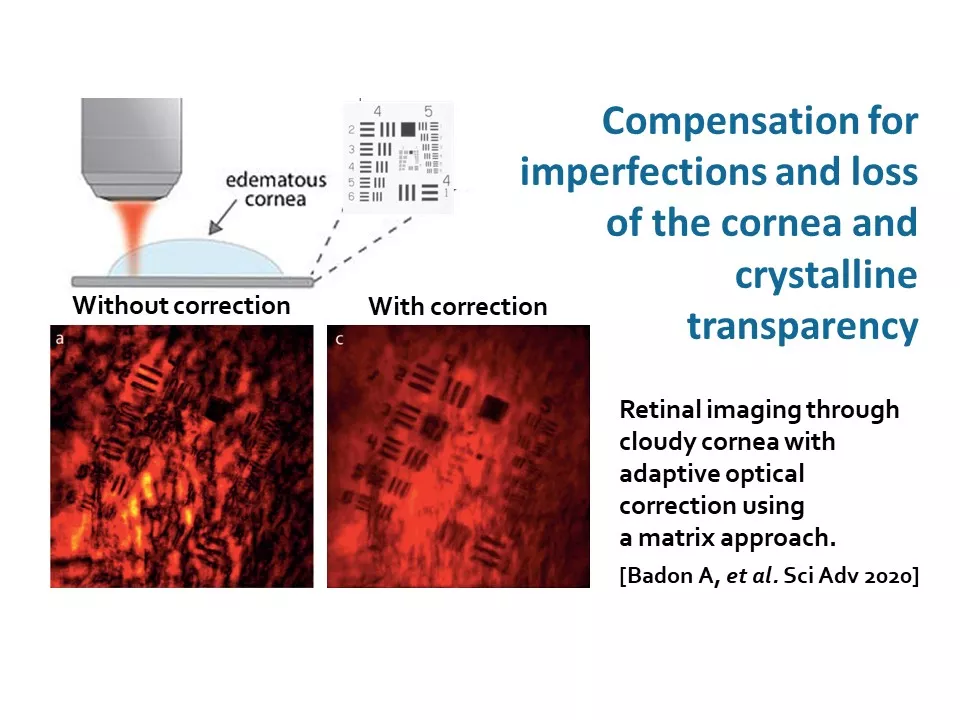
- The first focuses on overcoming human-factor limitations inherent to patient imaging. It includes (objective 1) the characterization of anterior-segment (the cornea and crystalline lens) transparency with tailored instrumentation and (objective 2) the compensation for imperfections and loss of transparency in the anterior segment.
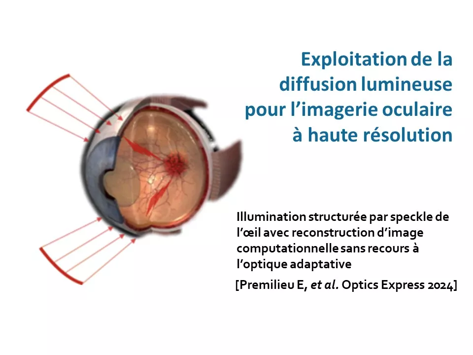
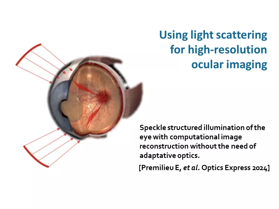
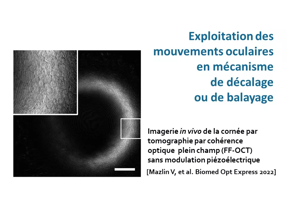
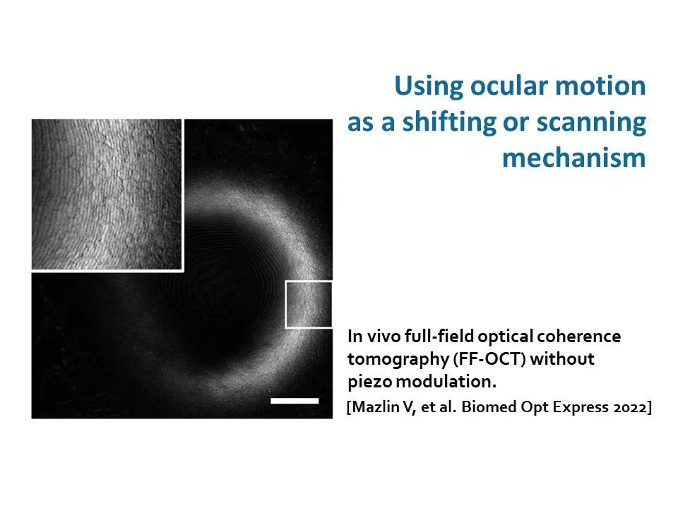
- The second line of research is exploring the possibility of turning some inherent limitations, such as eye movements and ocular scattering, into assets. The group is developing techniques to leverage (objective 3) light scattering for, and (objective 4) eye movements as a natural scanning mechanism in, high-resolution ocular imaging.
An inter-disciplinary approach
To achieve these objectives, an inter-disciplinary approach is employed involving a synergy between physicists/engineers and ophthalmologists, both within the group and at the national and international level.
Research areas
Standardized grading of corneal and lenticular opacities.
Retinal imaging through ocular opacities.
High resolution ocular imaging using scattering optics.
Full Field and curved-field optical coherence tomography (OCT).
Team members

Scientific publications
Below you will find the latest scientific publications in this field: Quantitative in vivo corneal and ocular microscopy.