


Even the retina recycles ; its night-vision circuits during the day
To process visual information, the retina makes the most of limited resources through a cellular wiring system that is still poorly understood. To investigate this, the teams led by Valentina Emiliani and Olivier Marre at the Institut de la Vision combined two-photon holography with large-scale recordings of retinal activity for the first time. Their findings, published on August 24, 2026, in the journal PNAS*, reveal a new principle of biological economy: the recycling of neural networks dedicated to night vision.
When light enters the eye, the retina immediately begins to process visual information. The results, in the form of electrical signals, are then sent to the brain by ganglion cells, the retina’s output neurons, whose long fibres form the optic nerve. Each ganglion cell "watches" only a small patch of the visual scene via a cluster of photoreceptors linked by horizontal, bypolar and amacrine cells, called its receptive field. This organization allows the retina to perform sophisticated computations with a limited number of cells. Such economy is a hallmark of the central nervous system: the brain’s total energy consumption is approximately 20 W, far less than the energy required to power a supercomputer. Yet, gram for gram, the retina consumes even more energy than the brain, making it one of the most energy-demanding tissues in the body. Part of its efficiency therefore lies in how its cells are wired together. Such performance is made possible by several principles built into the architecture of the receptive fields, which consists of a center and a surround (periphery) that influence each other.
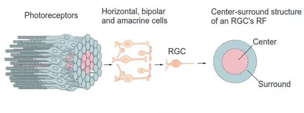
Scheme explaining ganglion cell’s receptive fields (modified from Principles of neural science. E. R. Kandel)
A key principle of retinal architecture is the ON/OFF dichotomy. Ganglion cells are found in two different types: ON cells respond when light increases in the center of their receptive field, and OFF cells when light decreases in the center of their receptive field. For some ganglion cells, when a light signal strikes the entire receptive field, its surround will attenuate or suppress the response triggered by the center. As a result, the ganglion cell responds more strongly to local contrast than to uniform light, which is particularly useful for detecting edges and movement.
Previous studies, however, revealed that many OFF ganglion cells also respond when light is on in their surround, the opposite of what they prefer in their center. This phenomenon, known as “antagonistic surround modulation” caught the attention of the teams led by Olivier Marre and Valentina Emiliani. Drawing on the Institut de la Vision’s expertise in circuit optogenetics, they developed a custom system combining optogenetics, two-photon holography and large-scale electrical recordings, a pairing never before used in the retina, to investigate this circuit in retinal samples.
Optogenetics involves genetically modifying neurons so that they produce light-sensitive proteins, allowing them to be activated remotely. A true revolution in neuroscience, it has made it possible to link different cell types to specific brain functions. The Institut de la Vision, thanks in particular to the work of Deniz Dalkara, possesses unique expertise in the design of customized viral vectors, vehicles capable of penetrating deep into the layers of the retina to deliver genetic information.
To dissect neural circuits with unparalleled spatial, temporal, and cellular resolution, Valentina Emiliani and her team use two-photon holography. This technology, which they have played a major role in developing, This technology, which they developed, shapes light in three dimensions to target individual cells in the retinal tissue with beams of light without affecting the surrounding cells.Using optogenetics and holography, the ganglion cells individually emit electrical signals. These signals are recorded by Olivier Marre and his team via a multi-electrode array (MEA) consisting of 252 electrodes arranged in a square slightly less than half a millimeter wide, placed in direct contact with the ganglion cells. These recordings first made it possible to map the receptive fields of up to hundreds of ganglion cells. The researchers could then record, with millisecond precision, how ganglions cells responded when individual cells upstream in the circuit between them and photoreceptors were switched on or off
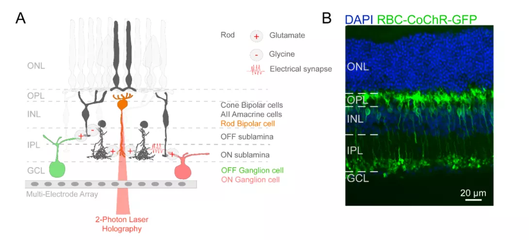
A. Schematic of the experimental protocol. Rod bipolar cells (RBCs) are optogenetically stimulated, activating a polysynaptic pathway involving AII amacrine and cone bipolar cells. Retinal ganglion cells integrate this signal, generating responses recorded using multi-electrode arrays (MEA). B. Retinal slice showing the expression of CoChR-GFP in rod bipolar cells.
Using optogenetics and holography, the ganglion cells individually emit electrical signals. These signals are recorded by Olivier Marre and his team via a multi-electrode array (MEA) consisting of 252 electrodes arranged in a square slightly less than half a millimeter wide, placed in direct contact with the ganglion cells. These recordings first made it possible to map the receptive fields of up to hundreds of ganglion cells. The researchers could then record, with millisecond precision, how ganglions cells responded when individual cells upstream in the circuit between them and photoreceptors were switched on or off.
Thanks to this unique system, which allows researchers to manipulate deep retinal circuits while directly measuring their impact on output neurons using only light stimulation, Valentina Emiliani and Olivier Marre identified a circuit that contributes to antagonistic surround modulation : the main pathway of night vision. Formed by rod bipolar cells and AII amacrine cells, this pathway make up a large fraction of the retina's neurons, yet they were thought to be saturated and so unusable during the day. Next, they showed how this pathway « piggybacks » the very cells that normally carry out classic surround suppression, inhibiting them to procduce the opposite effect.
In short, the same cellular network serves for different processing depending on the visual context. Thus, the retina "recycles" its internal wiring to optimize its biological resources rather than creating dedicated circuits for each mechanism. Similar forms of circuit reuse likely occur for other types of cells, both within the retina and in other regions of the brain. In fact, this is precisely why studying the retina is so valuable to neuroscience: its more accessible and easily manipulable circuitry allows us to understand the biological functioning of neural networks and the principles that govern these extremely complex tissues.This study is part of the European DEEPRETINA project, which aims to develop realistic models of the visual system's neural networks.
* G. Spampinato, F. Trapani, V. Calbiague-Garcia, T. Buffet, E. Orendorff, B.S. Sermet, G. Glaziou, D. Dalkara, E. Ronzitti, E. Papagiakoumou, V. Emiliani, & O. Marre, The rod bipolar cell pathway contributes to surround responses in OFF retinal ganglion cells, Proc. Natl. Acad. Sci. U.S.A. 123 (35) e2532792123, https://doi.org/10.1073/pnas.2532792123 (2026).