Live imaging: Patients and Cells
Kate Grieve’s team develops novel imaging technology to discover the health of cells in the patient’s living retina or in the lab.

Presentation
The two main technologies we explore are adaptive optics ophthalmoscopy and optical coherence tomography.
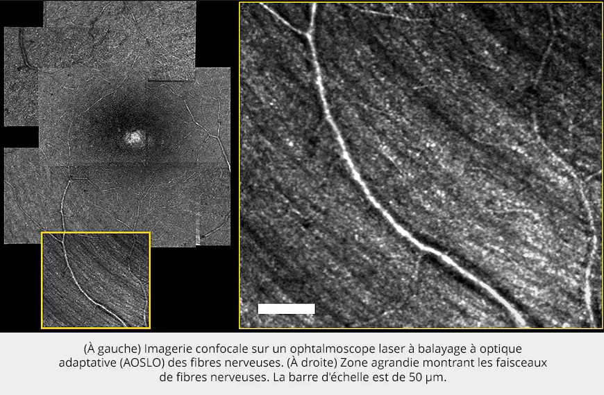
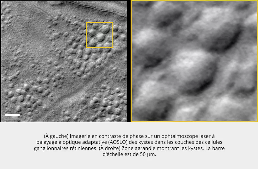
Imaging tools we developed in the clinic are used for diagnosis and follow up of cohorts of patients suffering from retinal disease. Advanced techniques such as phase contrast imaging and optoretinography for measurement of retinal function are the current focus of the group. We also develop full field optical coherence tomography to provide cellular resolution in the retina in a compact clinical device.
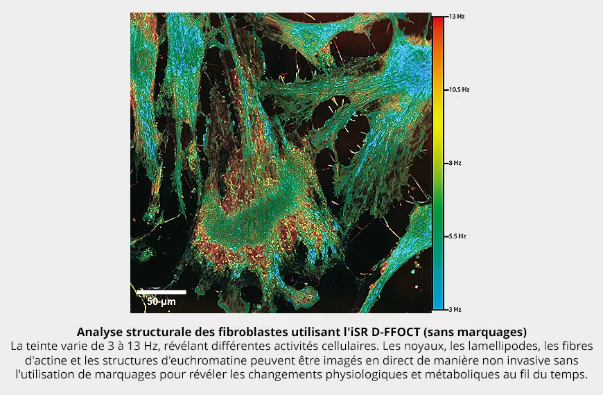
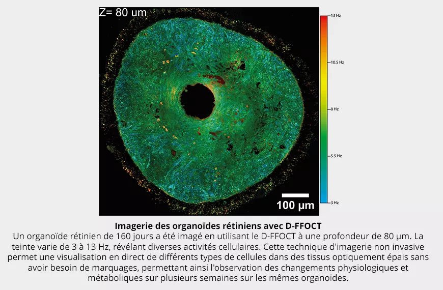
In the lab, we have adapted dynamic full field optical coherence tomography to allow non-invasive label free imaging of cellular activity in retinal organoids and cell cultures. Our dynamic imaging microscope allows 3D live imaging over seconds to month long periods to track developmental or degenerative processes and thus help to decipher disease origins.
Research areas
Adaptive optics
Optical coherence tomography
Retinal imaging
Live microscopy
Functional and structural imaging
Cellular resolution
Team members


Scientific publications
Below you will find the latest scientific publications in this field: Live imaging: Patients and Cells.