
Understanding vision
How do images form in our brain? The first thing we see is light, reflected back to us by the objects around us. It enters our eyes through the crystalline lens.
But how is light information captured, interpreted and transmitted? This film by Marc Desenne explains the anatomy of the eye, how it works and the different parts of the brain involved in vision.
Eye Anatomy
The human eye is a globe 2.2 to 2.5 centimeters in diameter that weighs between 7 and 8 grams. It is mobile thanks to 6 extraocular muscles. The eye is a high-performance organ, but also complex and fragile. It’s a sphere filled with liquid.
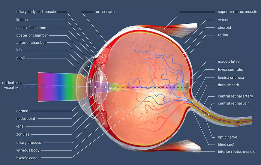
The 3 layers of the eye
Sclera (the outermost)
The sclera is a white, opaque layer that that constitutes five-sixths of the eyeball and consists of dense connective tissue fibers. The sclera protects the internal structures of the eye and allows the attachment of the extraocular muscles.
Choroid
The choroid is a well-vascularized and pigmented layer of the posterior part of the eye that is firmly attached to the fibrous tunic (sclera) by lamellae and rests on the nerve tunic (retina). It’s made up of two layers: the choriocapillaris and Bruch's membrane. The function of the choroid is to nourish the retinal cells.
Retina
The retina is the innermost layer of the eyeball, made up of two basic layers: the nerve retina (or the retina itself) and the retinal pigment epithelium. The function of the retina is to collect light stimuli, process them and transmit them to our brain.
The anterior chamber
The anterior chamber of the eye is composed of the cornea, the sclerocorneal limbus, the iris, the ciliary body and the ciliary muscles.
Cornea
The main function of the cornea is to focus light beams onto the lens. Due to its location and being the first structure that receives light and environmental components, the cornea is capable of detecting foreign bodies thanks to its rich innervation. Thanks to blinking, the corneal surface remains clean. The cornea is a highly innervated avascular element, which is why it is very sensitive. The cornea is also a transparent element, composed of 5 layers: anterior epithelium, Bowman's membrane, stroma, Descemet's membrane and posterior epithelium.
Sclerocorneal limbus
The sclerocorneal limbus is the transition zone between the transparent cornea and the opaque sclera (white of the eye). This area is highly vascularized which allows the cornea to be well nourished. The cells of the sclerocorneal limbus have a very high capacity for regeneration; they are undifferentiated cells (stem cells). The sclerocorneal limbus also allows the regulation of intraocular pressure thanks to the drainage system of the aqueous humor which it contains and which is located in the iridocorneal angle. Several canals lined with endothelia, known as the trabecular meshwork or spaces of Fontana, are found here. These converge and form the canal of Schlemm, which surrounds the cornea.
Iris
The iris regulates the entry of light into the eye. Inside is the pupil dilator muscle. It contains the dilator pupillae muscle, responsible for dilating the pupil under low light conditions (mydriasis), and the sphincter pupillae muscle, responsible for constricting the pupil under bright light conditions (miosis).It is melanin that determines the color of the iris. It all depends on the degree of production of this pigment. Brown eyes are therefore more pigmented than blue or green eyes. The iris is part of the vascular layer of the eye, also called the uvea. This layer is made up of the iris, the ciliary body and the choroid. It is the most anterior part of the vascular tunic or uvea, originates at the anterior limit of the ciliary body and is attached to the sclera 2 mm posterior to the sclerocorneal limbus. The central hole of this disc is the pupil.
The ciliary body
The ciliary body produces aqueous humor which helps regulate intraocular pressure, nourishes the structure of the eye and eliminates waste. It is the anterior extension of the choroid.
It has a triangular shape whose apex is the anterior limit of the retina and its base is located behind the iris. It is the support of the ciliary muscles and the ciliary processes. It has two parts: the pars plana, which serves as an insertion for the vitreous and the lens zone, and the pars plicata, which is located in the anterior part (they form an anterior vascular thickening of the choroid and another thickening which constitutes the ciliary process.
Ciliary muscles
The ciliary muscles participate in accommodation, that is to say focusing to see clearly up close (auto-focus). They are located at the level of the sclerocorneal limbus. Their fibers are distributed in three directions: meridian or longitudinal, radial or oblique, and circular or sphincteric.
What is the accommodation process in the eye?
Accommodation allows the human eye to see clearly at different distances. Its mechanism involves the contraction of the ciliary muscle, which induces a passive bulging of the lens thanks to the relaxation of its suspensory ligament (called zonule). It’s this bulging (increase in curvature) that allows the eye to reach the vergence necessary for close vision. Presbyopia occurs when accommodation is no longer sufficient to allow the eye to focus sufficiently up close.
Ciliary processes
Ciliary processes are extensions of the ciliary body with melanin granules inside and are covered by the ciliary epithelium. They produce aqueous humor, secrete and anchor the zonular (Zinn) fibers. Zinn fibers extend from the ciliary processes to insert into the lens capsule.
Lens
The lens provides a third of the total power of the eye, it allows accommodation to see clearly up close, and absorbs part of UV rays. It’s suspended all around by ligaments called zonule of zinn, attached to the ciliary muscles. The lens is normally transparent.
Aqueous humor
The aqueous humor is the transparent fluid which fills the front part of the eye between the cornea and the lens and which transports nourishing elements for these organs. The aqueous humor is constantly renewed by a mechanism of production and elimination, thus regulating intraocular pressure.
The ciliary bodies are responsible for producing aqueous humor. The aqueous humor has a circulation system within the eye. Aqueous humor maintains the shape of the anterior chamber structure. Its secretion and drainage help determine intraocular pressure.
Aqueous humor also helps nourish any parts of the eye without blood vessels, such as the back of the cornea and the front of the lens. Finally, the aqueous humor helps refraction of light entering the eye by channeling it towards the pupil then the lens.
The posterior chamber
Retina: macula and fovea
The retina is designed to receive light information and transforms it into interpretable signals for the brain. The retina is the sensitive layer of the eye where the image is formed. Its light-sensitive part is made up of cells called photoreceptors:
- Around 5 million cones : located mainly in the center of the retina. The cones allow fine visual acuity, definition of shapes and photopic vision. Cones allow color vision, green, red and blue cones.
- Around 120 million rods : located mainly on the periphery of the retina. The rods allow vision of movement and scotopic vision.
The retina is a thin, transparent membrane that lines the back of the eye and is made up of two parts:
- A central part with the macula, the fovea and the foveola which allow precision of vision (vision of details).
- A peripheral part which allows lateral vision, motion detection and vision in low light conditions.
Optic nerve
The essential role of the optic nerve is the transmission of information perceived by the retina to the brain. The information is first processed by the thalamus and then transmitted to the cerebral cortex. The optic nerve is a sensory nerve that allows the assimilation and integration of visual perception. Each optic nerve originates from retinal ganglion cells. It’s a cylindrical cord 5 cm long. This circular area, starting point of the optic nerve and passage of the vessels, is blind : it’s the optic disc, called the "blind spot". Two areas can be distinguished in the optic disc: the papillary cup and the neuroretinal ring.
Vitreous body
The role of the vitreous body is to support the rigidity and elasticity of the eyeball and keep the retina pressed against the wall of the eyeball. The vitreous body is a viscous element that liquefies with age, most of the volume of the eyeball is filled by the vitreous body.
The ocular adnexa
Eyelids
The main function of the eyelid is to protect the eyeball. The eyelid (one upper and one lower) is a fold of skin that covers the front of the eye. Its external face is covered with thin and elastic skin, its internal face is covered by the tarsal or palpebral conjunctiva. The internal structures of the eyelid are a series of muscles and a dense connective tissue structure called the eyelid tarsus, which houses the meibomian glands. In the skin of the eyelids there are sweat glands, fine hairs and sebaceous glands. The edges of the eyelids have eyelashes, which are stiff, curved hairs whose function is to protect the eye from the sun's rays and environmental particles.
Conjunctiva
The conjunctiva is a thin transparent mucous membrane that lines the posterior surface of the eyelid and the anterior part of the eyeball, over the sclera, around the cornea. The purpose of the conjunctiva is to protect the eye. It is responsible for maintaining the front surface of the human eye moist and lubricated, allowing the opening and closing of the eyelids without friction or irritation of the eyes. The conjunctiva protects the eye from external factors such as dust or microorganisms that may cause infections. The blood vessels of the conjunctiva help nourish the eye and the eyelids.
Lacrimal gland
The lacrimal gland is located in the lacrimal fossa, on the upper and outer surface of the orbit. It is a compound tubulo-alveolar serous gland, its alveolar units draining into the twelve lacrimal ducts, which pierce the conjunctiva, forming the lacrimal points. The tears go to the lacrimal ducts (upper and lower), which are located in the medial inner corner of the eye, forming the common lacrimal duct, which in turn flows into the lacrimal sinus, which joins the nasolacrimal duct. The upper eyelids transport tears to the anterior sclera and cornea, keeping them moist and protecting them from dehydration.
Eye muscles
The eye muscles, also known as the extraocular muscles, are responsible for eye movements when they receive signals from the brain via the optic nerve. Eye movements are carried out by six eye muscles: the lateral rectus, medial rectus, inferior rectus, superior rectus, inferior oblique and superior oblique. Inside the bony structure supporting the eye, all but one of these muscles - the inferior oblique - form a cone, with its tip directly behind the eyeball. This point is called Zinn's ring, and it is the point at which the optic nerve enters the eye.
Optic pathways
The optic pathways carry information from the retina, which has been stimulated by light, to the brain so that images can be correctly interpreted. The optic pathway begins with the stimulation of photoreceptors (rods and cones). Then, bipolar cells of the retina connect with ganglion cells, and the joining of the axons of the ganglion cells forms the optic nerve, which exits the posterior orbit to join the optic nerve on the opposite side to form the optic chiasm. At this stage, there is a crossing of nerve fibers. Nerve fibers leave the optic chiasm to reach the next station, which is the lateral geniculate body. The lateral geniculate body emits the optic radiation, which carries the information to the visual cortex of the brain (occipital region).