Development, evolution and function on commissural systems
Our team tries to understand how axonal guidance molecules regulate cellular interactions during the development of the visual system and in ocular diseases. We are also studying the function and evolution of commissural projections, particularly visual projections.
We are also taking part in the worldwide Human Cell Atlas (HCA), initiative, which aims to map all the cells in the human body. As part of this, we are coordinating the Inserm HuDeCA, consortium, which is looking at human embryonic development.

Presentation
The nervous system is made up of complex networks of excitable cells (neurons) connected by synapses. Neuronal connections are established during embryonic development. Neurons emit an extension, the axon, which grows in the brain towards target cells. The migration of neurons and the formation of a myelin sheath around the axon are other essential phases of neuronal development. For several decades, many researchers have been trying to discover the cellular and molecular mechanisms that control axon guidance during development. This work has shown that these processes have been highly conserved throughout the evolution of species. It has also been discovered that neuronal connections are plastic and that they are modified during normal physiological processes (particularly learning) but also in pathological situations, such as albinism.
Since its creation, our team has been studying the development of neuronal circuits in the nervous system. This area of research is expanding rapidly, as it has been shown that the mechanisms and molecules that regulate axon growth and neuron migration also modulate cellular interactions outside the brain, in many developing organs and in cancer tumours. The main aim of our work is to understand the function and mode of action of axon guidance molecules during the development of the nervous system, and in particular the visual system.
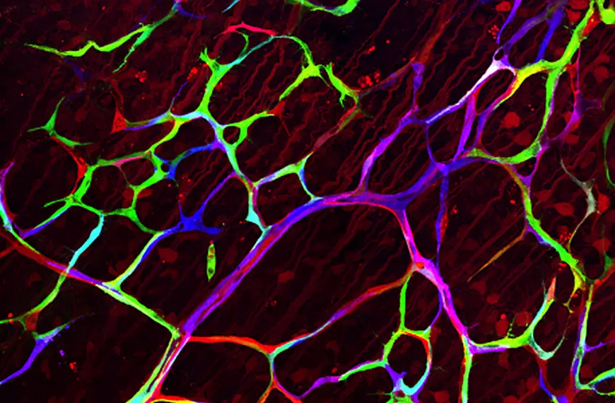
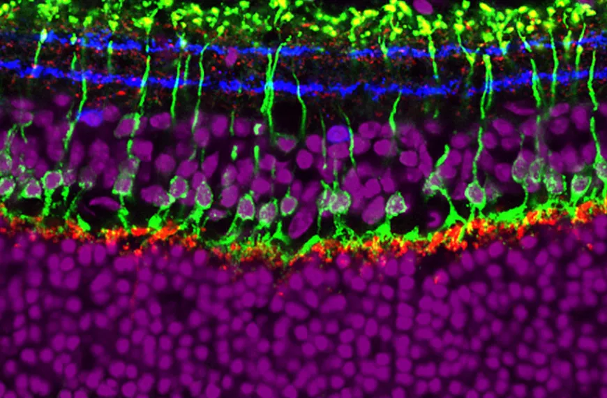
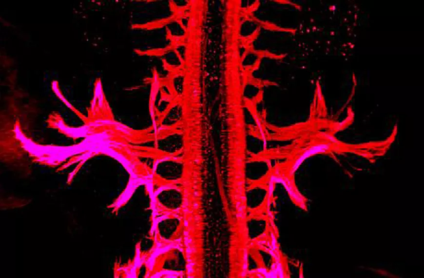
We are working on a limited number of models (retina, corneal nerves, pre-cerebellar neurons, commissural neurons). We are trying to understand how the cell layers in the retina are organised and how commissural axons can interconnect the two halves of the brain. To answer these questions, we use various animal models (birds, fish, rodents, etc.). We are trying to develop therapeutic tools to regenerate the cornea and the optic nerve.
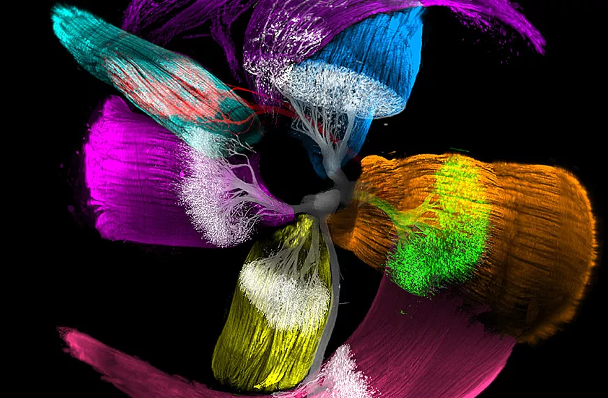
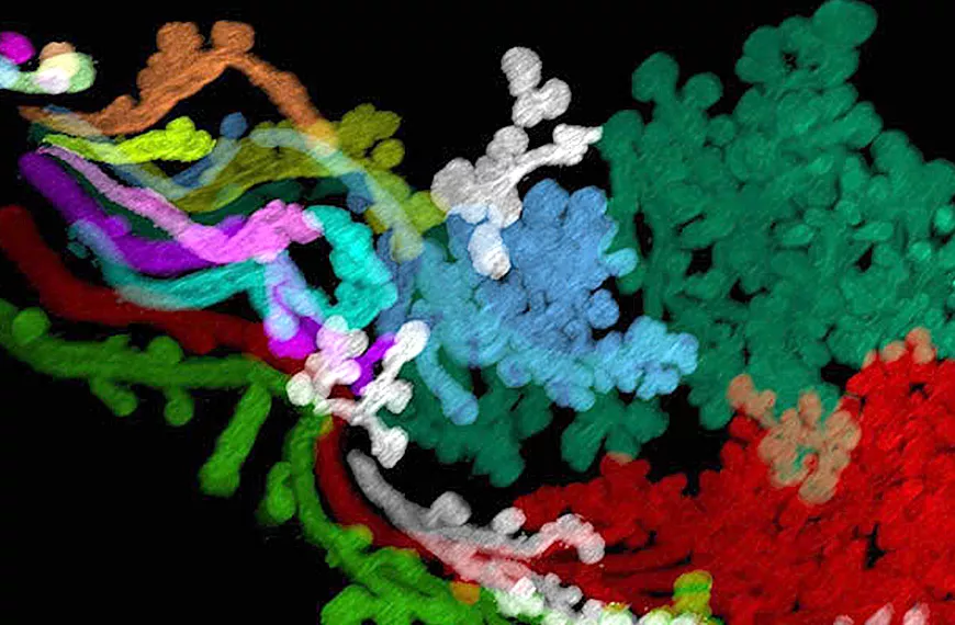
We are involved in a number of national and international initiatives aimed at applying modern techniques to the study of the human embryo in order to gain a better understanding of the origins of developmental diseases. For example, we are taking part in HuDeCA (Human Developmental Cell Atlas), a multi-year project initiated by Inserm to create a reference atlas of human embryonic and foetal cell types. One of our aims is to combine and integrate multiple technologies to establish the characteristics of cells in embryonic organs. Our team has developed an innovative 3D imaging method that makes it possible to visualise cells in organs that have been made transparent, using a microscope known as a light sheet microscope, which scans tissues using lasers. We recently published the first atlas of the development of the human head, and in particular the muscles that move the eyes.
Team members


Scientific publications
Below you will find the latest scientific publications in this field: Development, evolution and function on commissural systems.