
Mapping synapses in real-time
Researchers at the Institut de la Vision in Paris have developed a technique to map synaptic connections in the living brain with unprecedented precision and speed. Published in October 2025 in the journal Nature Neuroscience*, this breakthrough paves the way for a better understanding of the brain mechanisms underlying perception, learning and neurological diseases..
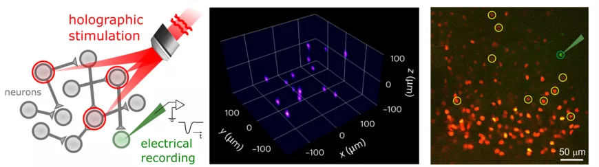
Left: diagram of the experimental approach to probe synaptic connectivity by optical stimulation of presynaptic neurons and electrical detection of postsynaptic neuron responses, as well as to analyze the presence and properties of synaptic connections.
Center: example of multi-point optical illumination for photostimulation of neural ensembles. Right: Example of a synaptic connectivity map in layers 2/3 of the primary visual cortex (V1) in mice. The postsynaptic pyramidal neuron (green) is electrically registered, while potential presynaptic neurons (red) are optically stimulated, individually or in groups. Neurons identified as connected are indicated in yellow.
A cubic millimeter of human brain contains hundreds of millions of synaptic connections, that allow neurons to exchange information. Essential to all our cognitive, perceptual and behavioural processes, these connections evolve over the course of a lifetime for example to adapt after an injury, through the phenomenon of brain plasticity. Understanding the organization of neural networks and their evolution is a major challenge in neuroscience, but synaptic connection mapping technologies did not provide information on an active brain. By combining several historically limited methods with state-of-the-art technologies, the Wavefront Engineering Microscopy team led by Valentina Emiliani at the Institut de la Vision has developed a new and less constraining approach, obtaining a map of neural networks through two photon holographic optogenetic.
An anthology of methods...
One of the oldest techniques for studying brain activity in a living organism is electrophysiology, implanting electrodes under the skull until insertion into a neuronal cell to record its electrical activity. Although very precise, this approach only allowed a very small number of neurons to be studied simultaneously, while being highly invasive. To circumvent these limitations, the researchers used two techniques pioneered by the Institut de la Vision.
The first is a specialty of Valentina Emiliani's team: holographic light shaping. Using liquid crystal devices, this technique allows the wavefront of a laser beam to be precisely modulated to generate a specific light pattern. By projecting this three-dimensional map onto a living tissue, it is possible to selectively illuminate one or more cells, to image them but also to activate them using a second technique: optogenetics.
Developed about twenty years ago, optogenetics uses gene therapy to allow cells to produce light-sensitive proteins, opsins. In the brain, this makes it possible to activate a neuron remotely with light.
The publication co-authored by Dimitrii Tanese and Valentina Emiliani combines these three methods to identify the synaptic connections of a neuron. First, they use optogenetics to modify pre-synaptic neurons, those that transmit information, to make them photosensitive. They then activate them using holography and measure the activity of the postsynaptic neuron, which receives the information, using an electrophysiology electrode. The presence of a synaptic response makes it possible to identify the existence of a connection and to characterize its nature as well as its strength.
But working on an active living organism required several innovations that make this publication an important step forward.
… Connected by innovations
To be able to activate neurons in a living brain, the researchers used two-photon holography: they project the pattern of the holographic map with infrared lasers capable of reaching cells deep into the brain tissue without affecting and damaging superficial layers. By using opsins adapted to two-photon stimulation, this approach makes precise 3D stimulation possible as well as repeated activation, which is essential for mapping synapses in a living brain.
Two-photon holography also eliminates the need for multiple electrodes, one per presynaptic cell, making connection mapping much faster and less invasive. To further reduce measurement time while maintaining high accuracy, the Institut de la Vision’s team collaborated with researchers at the University of Florida to develop a compressed sensing algorithm, a mathematical technique that allows a signal to be reconstructed from a limited number of measurements, more suitable for synapse mapping. Instead of stimulating presynaptic neurons one by one, the scientists stimulated them in groups of 5 to 10 neurons at a time, using holographic light patterns. Each pattern activates a different subset of neurons. With each stimulation, they measure the electrical response of the postsynaptic neuron via the electrode. This response is thus the sum of the contributions of connected presynaptic neurons in the stimulated group. Using compressed sensing, they unravel these responses to identify which presynaptic neurons are actually connected to the postsynaptic neuron.
Further progress ahead
For the first time, this system makes it possible to map a network of a hundred neurons in a few minutes while maintaining a cellular resolution, compared to several hours for conventional methods. Experiments conducted on mice, carried out by I-Wen Chen and Chung-Yuen Chan, co-first authors of the study, demonstrated the reliability of this technique, revealing patterns of connectivity in the visual cortex, a key region for the processing of sensory information.
In the longer term, the Vision Institute team is working to make this type of mapping completely non-invasive, by replacing the electrodes with fluorescent indicators. The team is developing two-photon holographic systems capable of detecting fluorescence and neuronal activity at depth – a step towards the complete optical mapping of brain circuits. This work was published in the journal Neuron, as well as an article on the Institut de la Vision's website.
*Chen IW*, Chan CY*, Navarro P, de Sars V, Ronzitti E, Oweiss K, Tanese D#, Emiliani V#. High-throughput synaptic connectivity mapping using in vivo two-photon holographic optogenetics and compressive sensing. Nat Neurosci. 2025 Oct; 28(10):2141-2153. DOI: 10.1038/S41593-025-02024-Y. Epub 2025 Sep 17. PMID: 40962966; PMCID: PMC12497651
Link to publication : Nature Neuroscience – DOI: 10.1038/s41593-025-02024-y