
The very first 3D mapping in the embryo
Enhancing our understanding of the development of complex structures that make up the human head and thus better understanding congenital anomalies causing malformations: this is the challenge that the team of researchers from Inserm, CNRS, and Sorbonne University at the Vision Institute, Claude Bernard Lyon 1 University, and the Hospices Civils de Lyon is on track to meet.
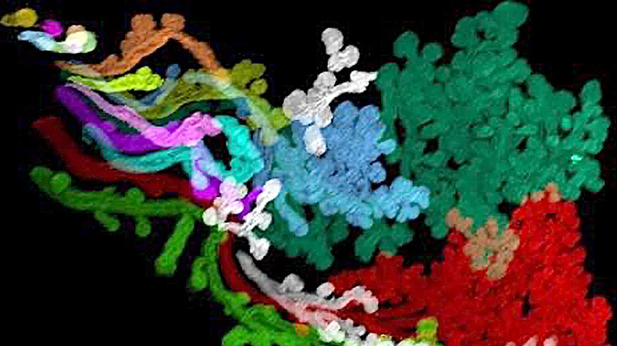
Thanks to an innovative technique that renders cranial structures transparent and then takes 3D photos of the cells composing them, this research team has been able to establish the very first three-dimensional map of the embryonic human head. These findings, published in Cell, have already helped to better understand how certain complex structures of the head, such as lacrimal and salivary glands or arteries of the head and neck, form. They pave the way for new tools to study embryonic development.
The head is the most complex structure of the human body. In addition to the muscles and skin that protect it, and the brain housed in the skull, it contains blood vessels, nerves, as well as endocrine glands (which secrete hormones directly into the bloodstream), such as the pituitary gland, and exocrine glands (which secrete substances to the exterior), such as salivary glands, which produce saliva, or lacrimal glands, which secrete tears.
Current knowledge about the development of the human head and its complex structures is rudimentary and comes mostly from studies conducted in the first half of the 20th century, using simple histological sections. Thus, although head malformations exist in about a third of babies with congenital anomalies, the mechanisms that control the development of the human head are still poorly understood.
The research team led by Alain Chédotal, Inserm research director at the Vision Institute (Inserm/CNRS/Sorbonne University) and professor at the MéLiS laboratory of Mechanisms in Integrative Life Sciences (Inserm/CNRS/Claude Bernard Lyon 1 University/Hospices Civils de Lyon), and by Yorick Gitton, CNRS research fellow also at the Vision Institute, used an innovative microscopy method to shed new light on the development of the human head.
The technology implemented had previously been used on embryos by the team to study the development of other human organs. It is called "transparentization" because it allows organs to become transparent to light. The transparentized sample is then imaged in 3D using a special microscope that scans with a thin sheet of laser light. This allows locating in situ the cells that constitute embryonic tissues.
The researchers succeeded in applying this technique to embryos at different stages of development, drawn from the human tissue biobank established as part of the HuDeCA (Human Developmental Cell Atlas) program coordinated by Inserm. Thanks to the obtained images, they were able to create the first three-dimensional map of the embryonic human head.
In a second phase, the research team used virtual reality to analyse the 3D images and virtually 'navigate' through the embryos. This enabled us to discover previously unknown characteristics of the development of cranial muscles, nerves and blood vessels, the skull and cranial exocrine glands," explains Alain Chédotal. For example, it had never been possible to study the very early stages of development of the salivary and lacrimal glands in humans. Our work has enabled us to begin to visualise and better understand the mechanisms behind the development of these extremely complex anatomical structures", he adds.
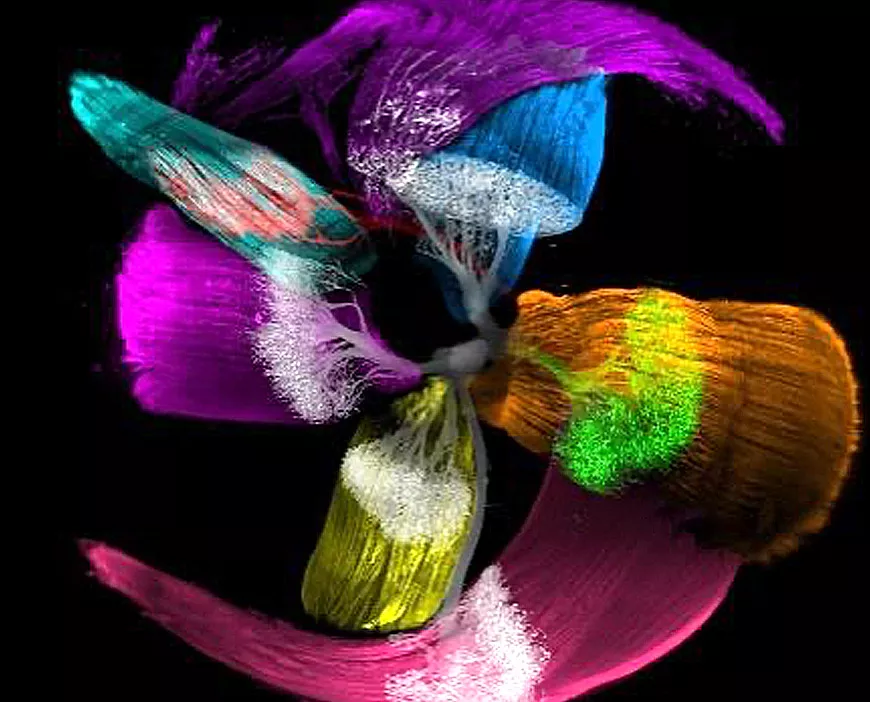
The scientists also set up a web interface (Hudeca.com) allowing not only access to the images obtained in this work but also to models for 3D printing and interactive 3D reconstructions of human embryos. This platform provides valuable resources that can also contribute to the training of medical students.
In future work, the research team will attempt to map all the cells of certain organs, such as the retina. "At this stage, it's a bit like if we had mapped the continents and countries but still needed to position the cities and inhabitants," explains Alain Chédotal, whose team will also collaborate with doctors to apply the technology to pathological samples. "The new knowledge about human embryology provided by this work, as well as the new tools developed, have important implications for understanding craniofacial malformations and neurological disorders, as well as for improving diagnostic and therapeutic strategies," concludes the researcher.
[1] Launched in 2019, the cross-cutting HuDeCa program led by Inserm aims to build the first atlas of cells of the human embryo and fetus. It also aims to structure research in human embryology at the French level and to develop databases. In the longer term, this program should serve as a basis for understanding the origin of chronic diseases or congenital malformations. See the press release dated March 23, 2017, on this subject: https://presse.inserm.fr/lembryon-humain-comme-vous-ne-lavez-jamais-vu/27807/
[2] With the specific exception of the brain, which is not a structure studied in these works.
A tridimensional atlas of the developing human head. Raphael Blain1, Gérard Couly1, Eimad Shotar1, 2, Joséphine Blévinal1, Maryne Toupin3, Anais Favre1, Ali Abjaghou1, Megumi Inoue1, Edwin Hernández-Garzón1, Frédéric Clarençon2, Frédéric Chalmel3, Séverine Mazaud-Guittot3, Paolo Giacobini4, Yorick Gitton1, * and Alain Chédotal1, 5, 6, - Cell : https://doi.org/10.1016/j.cell.2023.11.013
Sources :
- 1 Sorbonne Université, Inserm, CNRS, Institut de la Vision, Paris, France
- 2 Department of Interventional Neuroradiology, Pitié-Salpêtrière Hospital, Paris, France, Sorbonne Université, Paris, France
- 3 Inserm, EHESP, Univ Rennes, Irset (Institut de recherche en santé, environnement et travail), UMR_S 1085, Rennes, France
- 4 Univ. Lille, Inserm, CHU Lille, U1172 - LilNCog - Lille Neuroscience & Cognition, LilleF-59000, France
- 5 Institut de pathologie, groupe hospitalier Est, hospices civils de Lyon, Lyon, France
- 6 University Claude Bernard Lyon 1, MeLiS, CNRS UMR5284, Inserm U1314, 69008, Lyon, France