
Proof of concept for a new subretinal implant
A new strategy for restoring vision has been proven to work, and the Institut de la Vision has played a key role in this. Engineers Antoine Chaffiol and Corentin Joffrois have successfully tested a subretinal photovoltaic implant currently under development, the results of which were published last January in Science Advances.
Before reaching the clinic, retinal implants under development must go through many validation phases, including proof of concept. This first stage, which evaluates the effectiveness of the technology for its intended application, is essential in the process of designing and evolving towards translational applications. Recently, Antoine Chaffiol and Corentin Joffrois, research engineers at the Institut de la Vision, validated the experimental concept of a new photovoltaic subretinal implant designed and manufactured in Istanbul by Sedat Nizamoglu's team at Koç University. By placing the device under an ex-vivo retina, the two engineers showed that it was possible to effectively stimulate retinal ganglion cells (cells connected to the optic nerve) by using light intensities compatible with use in patients.
Restoring vision with implants: a recognized strategy
In a retina, the processing of light information begins at the level of the light-sensitive cells, called photoreceptors. Visual information is translated into electrochemical signals that are then transmitted to ganglion cells, via several layers of cells, and then to the brain via the optic nerve. The degeneration of the light-sensitive cells of the retina is one of the leading causes of vision loss worldwide. But in cases where only the first cell layer is impacted, research has shown that it is possible to restore some light information circulation by stimulating the remaining cells with an implant. Placed under the retina, these visual restoration devices are designed to replace non-functional photoreceptors by converting a light stimulus into an electrical stimulus, such as the PRIMA photovoltaic implant, or a mechanical stimulus, such as the photoacoustic implant.
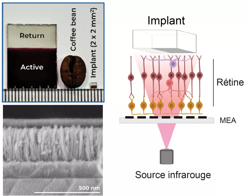
In this study, the team from Koç University is interested in a new type of photovoltaic implant. It is composed of an active part (the brown part in the photo), which consists of a layer of photoabsorbing nanocrystalline material interpenetrated with semiconductor nanowires, all less than 500 nm thick. This layer generates an electric current under infrared lighting: the light provides the energy needed to remove electrons that will migrate into the implant. This charge displacement generates a potential difference and therefore the appearance of an electrical voltage. As nanowires facilitate the migration of charges, their length has been the factor in optimizing the light-to-electric current conversion efficiency.
After manufacturing, the question of the functionality of the implant arises: is it possible to stimulate retinal cells with it? It is on this crucial step that the electrophysiology know-how of the two researchers came into play.
Proof of concept by electrophysiology
To test the proper functionality of the implant, the two researchers from the Institut de la Vision checked ex-vivo the activation of the ganglion cells of a blind rodent retina by measuring their electrical activity, this is called electrophysiology. To do this, they placed the retina on a MEA device or MultiElectrode Array device.
"It's like a microscopic fakir carpet with lots of little spikes, except it's not a fakir on it but a retina." Antoine Chaffiol illustrates, "Each small recording site - there are 256 - is in contact with the part of the retina that generates action potentials, in this case it is the ganglion cells."
The implant is positioned in place of the defective photoreceptors, on the part of the retina that is not in contact with the MEA device. It is then illuminated with an infrared laser pulse, for only a few milliseconds.
"The information [from the stimulus] is transmitted through all the layers of neurons in the retina to the ganglion cells that are recorded on the MEA. " explains the researcher. "We have thus succeeded in recording rapid, sharp and repeatable activations of the ganglion cells with infrared flashes that are very short and at intensities compatible with clinical safety standards. The responses were spatially well-defined and not artefactual, as we have been able to show with manipulations using controls."
On the strength of these initial results, obtained thanks to the experimental knowledge of the Institut de la Vision, the Sedat Nizamoglu research team wants to continue developing the prototype to make it more flexible in order to fit the curved shape of the retina in vivo.
The Institut de la Vision at the heart of implant development
This miniature solar panel is promising but before clinical application, further experimental validation steps are required in-vivo.
"With a version that can be implanted under the retina, we will soon be able to make in vivo cortical recordings directly in the visual cortex to see if the information is transmitted there and to be able to characterize it." Antoine Chaffiol explains, "But first there is a technical challenge to be met so that the implant is flexible, porous, biocompatible and of course still functional. The first flexible prototype will arrive very soon."
This international collaboration, within the framework of the ERC MESHOPTO obtained by Professor Sedat Nizamoglu in association with Serge Picaud, highlights the know-how of the Institut de la Vision's researchers in the functional analysis of visual restoration strategies, as well as their involvement in the development of tomorrow's retinal implants.
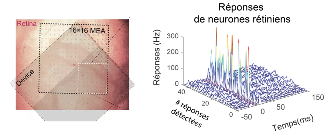
Left: infrared image of a rodent retina positioned on the MEA (mini-electrode array), outlined by the black dotted square, with the implant placed above it (in grey);
Right: responses from retinal neurons (detected within the white square on the left) stimulated by the photovoltaic implant following a 10 ms (milliseconds or thousandths of a second) infrared light stimulus. The brief responses, known as ‘spikes’, lasting less than 10 ms, are measured by the electrodes approximately 16 ms after the light stimulation
Scientific article: Tarik S. Kaya et al. Photovoltaic nanoassembly of nanowire arrays sensitized with colloidal nanocrystals for near-infrared retina photostimulation. Sci. Adv. 12, eaea7001(2026). DOI:10.1126/sciadv.aea7001