
A New Milestone in In Vivo Corneal Imaging
Researchers on Alain Chédotal’s team have developed a protocol that allows them to track the development of corneal nerves in living mice over nearly the entire lifespan of the animal. Published* in June in The Journal of Neuroscience, their findings show that the architecture of this neural network is not static: certain superficial fibers are constantly reorganizing, while deeper nerves remain remarkably stable. This approach opens up new avenues for studying the effects of injury, aging, or therapeutic interventions on corneal innervation.
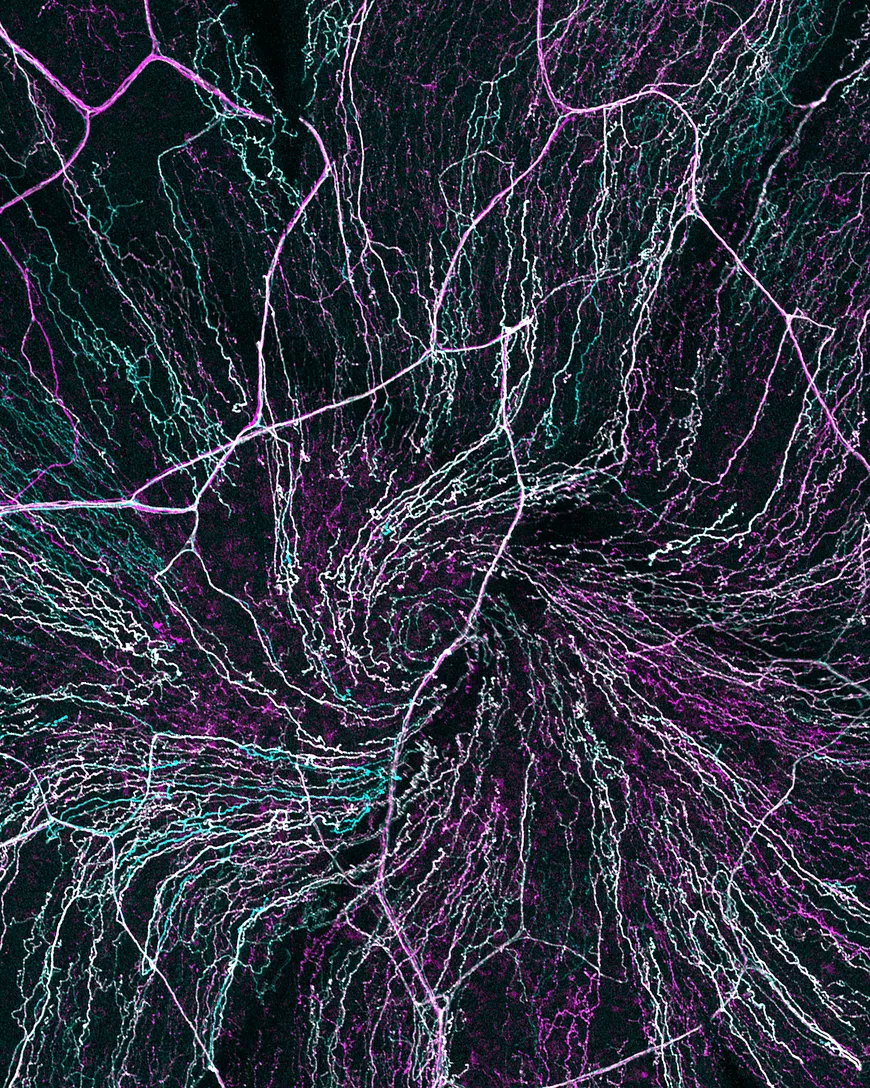
A mouse cornea, innervated by sensory nerves originating in the brain. This image shows neuronal extensions, or axons, forming the characteristic vortex (spiral). Anti-βIII tubulin (magenta) labels all axons, and those specifically involved in pain processing express GFP (cyan). Elena Bizzarri and her colleagues used spinning-disk confocal microscopy to track how individual axons in the cornea develop, mature, and remodel over time.
Credits: Quentin Rappeneau and Alain Chédotal
The cornea, a vital interface between the external environment and the internal eye tissues, is an amazing structure. This transparent window, which protects the eye and directs light toward the lens, is not inert—quite the contrary. The cornea is the most densely innervated tissue in the human body, with 300 to 400 times more nerve endings than the skin. This high nerve density ensures its structural integrity and facilitates healing. It also triggers tear secretion and the eyelid blinking reflex, both of which are essential for eye health. Despite their importance, our understanding of the development, maturation, and remodeling of corneal nerves remains limited.
An Approach That Preserves the Animal
Most studies conducted on mouse models of corneal diseases rely on large cohorts of mice from which samples are collected ex vivo. However, these approaches do not allow for tracking the evolution of each cornea over time within a single animal, which complicates the interpretation of experiments, particularly when testing the effects of pharmacological agents. In contrast, in vivo imaging allows researchers to study a single individual’s long-term response to a treatment or surgery. This type of imaging not only improves the interpretability of the data but also significantly reduces the number of animals required for the study, in accordance with the 3Rs principle (replace, reduce, refine).Until now, in vivo imaging of neural connections in animals was not fast or precise enough to track their subtle changes. It was also limited to relatively short time frames, generally not exceeding a few weeks. Alain Chédotal’s team has developed a protocol that allows for regular observation of corneal nerves over the course of months, and up to 2 years—the lifespan of a mouse. The researchers used transgenic mice whose corneal nerves express fluorescent proteins.
To perform the imaging, the researchers use a high-resolution spinning-disk microscope. This device consists of two disks spinning at several thousand revolutions per minute. The first disk, composed of microlenses, splits an incident light beam into hundreds of secondary beams, which illuminate the sample. In this case, the mice are transgenic mice whose corneal nerves express fluorescent proteins. These proteins react to the light beams and, in turn, emit light, which is collected through the holes in the second disk before being detected by a highly sensitive camera. This technique allows for the rapid acquisition of high-precision images with minimal noise (light interference) and at a light intensity low enough to limit phototoxicity and potential overheating caused by the laser beams.
Incoming light is focused onto the cornea without direct contact, using immersion objectives and an aqueous ophthalmic gel as an intermediary. This allows researchers to observe neural networks within the transparent cornea without resorting to external dyes or invasive surgical procedures. By tracking the same mice throughout the study, rather than comparing different animals, researchers can reduce the number of mice needed for their project and also eliminate some of the biases associated with individual variability. A control group of mice imaged less frequently confirmed the safety of this protocol.

To isolate individual axons within the dense network provided by the microscope, the researchers use the syGlass virtual reality software, which enables precise manual segmentation in three dimensions.
The Cornea, a Promising Model
Using this system, Alain Chédotal and his team have conducted the longest in vivo imaging study to date on the neural dynamics of an animal. The researchers tracked and individually quantified, over time, the fate of nerves—including their growth, retraction, and remodeling—during postnatal development and aging. They also monitored axonal regeneration in real time following a lesion of the corneal epithelium.
Their results indicate that axons on the surface of the cornea are constantly rebuilding and retracting throughout life, unlike those deeper within the cornea, which serve as a stable structural framework. Over time, each individual pathway within the corneal nerve network is unique, though there is a consistent trend toward decreasing density with age. The best illustration of this phenomenon is the shifting of the spiral organization of the network formed by nerve fibers located in the outermost layers of the cornea.
Although long described as a characteristic anatomical landmark, this vortex appears here as a surprisingly unstable structure, whose position and shape drift over the course of months or even disappear.
Researchers at the Institut de la Vision have thus developed a high-resolution imaging platform to track changes in the corneal nervous network over time. This technique, designed to be as minimally invasive as possible, allows them to monitor the development, maintenance, and degeneration of axons in the corneas of rodents. These results provide a baseline for understanding alterations in corneal innervation in conditions such as refractive surgery, dry eye syndrome, neurotrophic keratitis, sensory neuropathies, and corneal pain. The new perspectives offered by this protocol make the cornea—a structure that is accessible to imaging and densely innervated—a particularly interesting model. It could become a prime area of study for investigating the mechanisms of peripheral nervous system plasticity in response to injury, aging, and therapeutic interventions, beyond the scope of ocular pathologies.
*Analysis of Corneal Axons Remodeling In Vivo during Development and Aging Using Long-Term Imaging in Mice,