

ERC-PoC: towards new tools for ophthalmic diagnosis
Kate Grieve, a research director at the Institut de la Vision, has been awarded one of the European Research Council’s (ERC) prestigious Proof of Concept (PoC) grants for her FFORGE project. Its aim is to bring the optoretinography technology developed in a close collaboration between Pedro Mecê’s team at Institut Langevin and Kate Grieve's team at the Institut de la Vision, closer to clinical use, in particular to facilitate the diagnosis of age-related eye diseases such as AMD.
The FFORGE project aims to transform ophthalmic diagnosis by moving from structural imaging – which merely shows the anatomy of cells – to functional imaging capable of measuring their activity. Whilst doctors can now count retinal cells at the back of a patient’s eye, the examination does not always make it possible to determine whether these cells are responding correctly to light. By focusing on Age-Related Macular Degeneration (AMD), the team hopes to detect early functional deterioration, occurring well before cell death or irreversible structural changes. This project therefore aims to provide clinicians with a robust, rapid and non-invasive tool for the day-to-day monitoring of patients..
10 years of fundamental and translational research
The FFORGE project builds on a long-standing collaboration between the Institut de la Vision and the Institut Langevin, which specialises in imaging and wave physics. As early as 2014, Professors José-Alain Sahel and Mathias Fink jointly secured European funding (ERC Synergy HELMHOLTZ) to develop and translate imaging technologies into clinical practice, one of which was full-field optical coherence tomography (FFOCT). This work led to the creation of the start-up SharpEye by Kate Grieve and her colleagues at the Institut de la Vision and the Institut Langevin.
Appointed Inserm Research Director in 2021 and leading her own team, ‘Live Imaging: Patients and Cells’, Kate Grieve has secured further European funding: the ERC Consolidator Grant ‘Optoretina’. This project involves combining several optical methods to develop new tools for the functional analysis of the retina. Kate’s innovations quickly led to her first ERC Proof-of-Concept (PoC) grant: LiveORG. A start-up, Lutèce Dynamics, was founded to commercialise their dynamic cell imaging techniques – particularly for organoids – for use in research. However, the researcher has not neglected the clinical side of her work. With the support of the Langevin Institute, SharpEye and the Quinze-Vingts Hospital, she continues to develop systems capable of non-invasively measuring the functionality of retinal cells directly within the patient’s eye. The ERC PoC grant for the FFORGE project recognises this work by providing the resources needed to realise its ambition of facilitating the diagnosis of age-related eye diseases.
Greater comfort for the patient, greater precision for the doctor
Until now, images of the eye obtained using FFOCT have made it possible to count retinal cells, but not to determine whether they respond to light. To obtain this information, a recent technique is becoming increasingly widespread amongst doctors: optoretinography (ORG). The principle behind it is to detect micro-changes in the structure of photoreceptors when they are exposed to a flash of light: they contract and expand by just a few tens of nanometres (one millionth of a millimetre). This change is so minute that it escapes detection by conventional optical imaging systems. Whilst ORG shows great promise, current systems are complex to use, making the examination time-consuming and uncomfortable for patients. Once the data has been collected, it takes several hours to process and produces a set of curves that are difficult for the doctor to interpret.
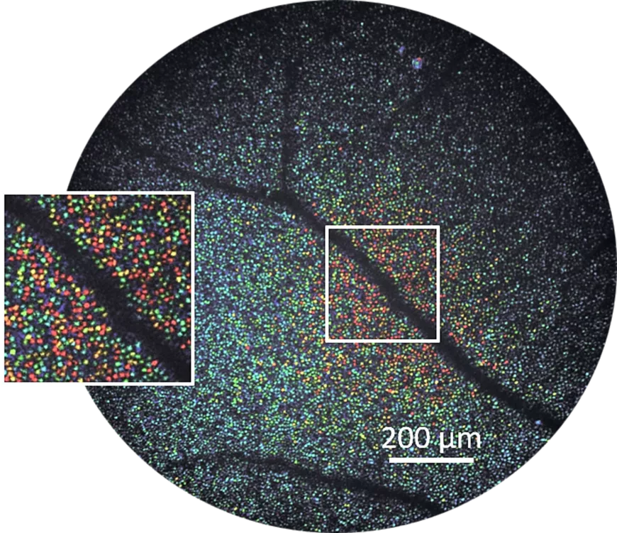
Kate Grieve and her project collaborators, Maxime Bertrand (SharpEye), Pedro Mecê (Institut Langevin) et Michel Paques (Hôpital national des 15-20) have chosen to develop a new OCT method to provide doctors with a functional map of a patient’s eye in a matter of seconds. To achieve this, the researchers are utilising the physics of light waves to capture information on the dynamics of photoreceptors. They are therefore combining FFOCT with a system for shaping the light source’s spectrum. By ‘sculpting’ the light spectrum using a filter, they are able to adjust the depth of the tissue slice being observed to detect light reflected from the various structures of interest. This process transforms even the slightest nanometric movement into a detectable variation in light intensity, enabling the cellular response to be visualised as a flicker on the image, almost in real time. Data acquisition takes just five seconds, and processing takes a few tens of seconds thanks to algorithms optimised by SharpEye. The end result is an intuitive colour map that quantifies the functionality of the retina’s photoreceptors, offering not only immediate diagnostic assistance to the clinician but also greater comfort for the patient.
This unique synergy between wave physics and clinical ophthalmology has already been shown in the literature to be useful in the study of rare retinal diseases, such as retinitis pigmentosa. The aim of the PoC is to extend this to other diseases, particularly those linked to ageing, such as AMD. Scientific literature shows that a decline in photoreceptor function occurs before cell death. Detecting this decline at an early stage would pave the way for better management, but also accelerate research by identifying associated biomarkers in collaboration with doctors.